Bone fractures are the bane of people with unhealthy or fragile bones. That is especially true for the elderly, for whom a fracture that occurs spontaneously or as the result of a fall or other trauma can be life-threatening. However, it has been difficult for healthcare providers to measure bone health accurately. With the advent of an ingenious, game-changing technology called microindentation, that has changed.
Most fragility fractures occur in patients whose bone density (“DXA”) scans do not show osteoporosis, which develops when bone mineral density and bone mass decrease. In fact, these scans often miss the effects of many circumstances that negatively affect bone health, such as disease, drug treatment, lifestyle and age.
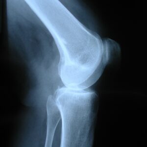
Strong, healthy bones require good structure, adequate mineral density and good-quality bone tissue. Until recently, bone health could be assessed only through indirect imaging methods like bone-density scans, which use a small dose of ionizing radiation to produce images of bones inside the body — usually the lower (or lumbar) spine and hips — to measure bone loss. It is the most common method for diagnosing osteoporosis, which reflects an individual’s risk for developing osteoporotic fractures.
However, although bone-density scans measure the amount of bone tissue, they do not provide information about how well the tissue is functioning or the quality of the tissue material. Other imaging techniques, such as CT or MRI scans, offer insight into the structure but fail to quantify tissue function and quality.
Tissue quality is a composite of properties that enable bone to resist fracture, such as its microarchitecture, accumulated microscopic damage, the quality of collagen, mineral crystal size, and bone turnover. Until recently, we had no technology that could provide a reliable quantitative measure of those qualities. Instead, we have had to rely on other factors like medical and family history to estimate a patient’s bone health.
But now we have an excellent new tool. BoneScore is a direct approach to evaluate bone tissue safely and directly, and it is an in-office procedure. A local anesthetic is used to numb the measurement site. Then a sterile acupuncture-like tip is slipped through the skin to the surface of the tibia, the “shinbone,” where the practitioner takes several readings.
After the readings, the BoneScore (called the Bone Material Strength index) is displayed. Higher scores reflect greater resistance to the indentation challenge (i.e., that the bone is harder) and lower scores reflect less resistance (i.e., softer bone). The first procedure of its kind, it is useful not only as a diagnostic tool to determine bone health but also as a research tool for measuring the effects of various interventions — such as drugs, diet or exercise.
Several years ago, a group of academic researchers in the Netherlands conducted a systematic review of all clinical studies using this approach. Their analysis included 38 studies, which showed that the technique was valuable — that it “can identify patients with primary osteoporosis and fractures, patients with secondary osteoporosis due to various underlying systemic disorders, and … that this tool can detect changes in bone material strength index (BMSi), following bone-modifying therapy including use of corticosteroids.”
BoneScore and similar devices are extremely useful and will become even more so with additional experience and studies. In the years after the Dutch researchers’ publication, the BoneScore medical device received FDA clearance, the American Medical Association established an exploratory reimbursement code, and the NIH funded additional studies.
This is the stuff of real medical progress, especially for older Americans.